For decades, textbooks have taught that the human brain emerges from a single pool of early cells, but a new Stanford Medicine study upends that assumption by showing the organ actually develops from two separate cellular lineages that merged over evolutionary time. The finding creates a clear split between the forebrain, which handles language and abstract thought, and the hindbrain, which governs breathing, heartbeat and other automatic functions.
The research, led by associate professor Kyle Loh and published in Nature Neuroscience, used single‑cell sequencing and lineage‑tracing techniques to map the developmental trajectories of human embryos. The data reveal that progenitor cells destined for the hindbrain diverge from those that form the forebrain and midbrain far earlier than previously believed. This structural insight rewrites the classic neurodevelopmental map and explains why scientists have struggled for years to grow hindbrain neurons in the laboratory.
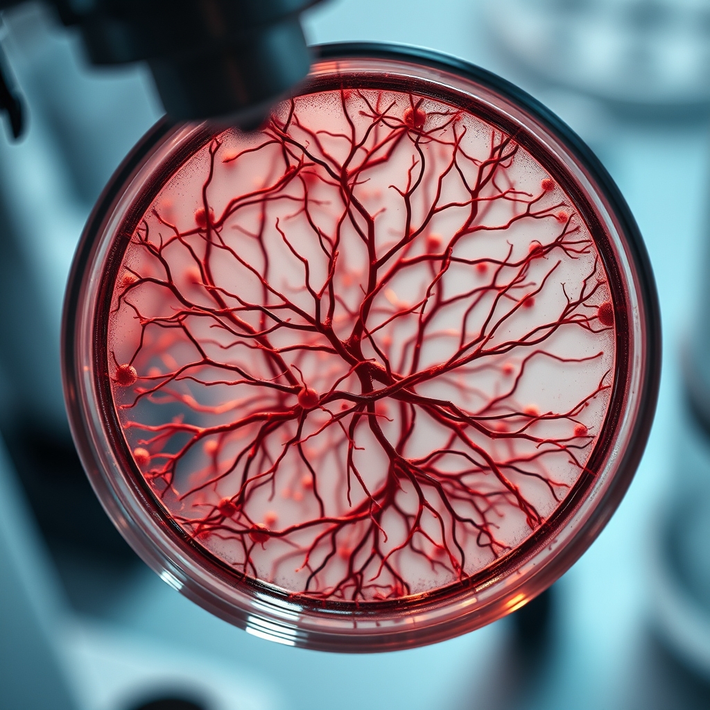
Why does this matter? The hindbrain houses the brainstem, a region implicated in life‑threatening disorders such as amyotrophic lateral sclerosis (ALS) and spinal muscular atrophy (SMA). By isolating the specific progenitor population that gives rise to hindbrain neurons, the Stanford team succeeded in cultivating these cells in petri dishes for the first time. "We've shown for the first time that the front of the brain arises from a totally different progenitor cell than the back of the brain," Loh said. "Our discovery means that we can now grow neurons from the back of the brain, the hindbrain, in a petri dish and study their functions."
This breakthrough opens a practical pathway for disease modeling. Researchers can now generate patient‑derived hindbrain neurons to test drug candidates for ALS, where degeneration affects both forebrain and hindbrain circuits, and for SMA, the leading genetic cause of infant mortality. The ability to study these cells in vitro shortens the gap between animal models and human pathology, potentially accelerating therapeutic development.
Beyond the immediate biomedical implications, the two‑origin model aligns with a broader trend in biology: recognizing that many complex organs are composites of older, evolutionarily distinct modules. Similar dual‑origin concepts have emerged in immunology and developmental genetics, reflecting a shift toward modular thinking in life‑science research. This perspective may influence how future technologies—such as organ‑on‑a‑chip platforms and AI‑driven developmental simulations—are designed, emphasizing the need to respect distinct lineage pathways.
Clinicians, biotech investors, and policy makers should note that the discovery could reshape funding priorities. Grants that previously bundled all brain research under a single umbrella may now be split to support targeted hindbrain studies, especially for rare neurodegenerative diseases. Moreover, pharmaceutical pipelines that have struggled with brain‑stem toxicity can benefit from more accurate cellular models, reducing costly late‑stage failures.
In practical terms, the study also explains a long‑standing puzzle in neuroscience: why attempts to grow brainstem cells repeatedly failed. The answer lies in the mismatched developmental cues used in earlier protocols, which assumed a uniform progenitor source. By providing a lineage‑specific recipe, Stanford’s work paves the way for reproducible, scalable production of hindbrain neurons.
As the field digests this paradigm, the next steps will likely involve integrating the two developmental maps into a unified atlas of human brain development. Such an atlas would serve as a reference for both basic scientists and clinicians, ensuring that future research builds on a more accurate foundation.